![]() Figure 3 of
Clarke, Mol Vis 4:3, 1998.
Figure 3 of
Clarke, Mol Vis 4:3, 1998.
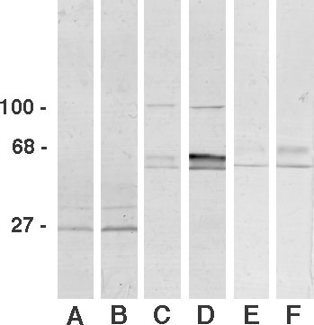
Figure 3. Immunoblots from Normal and Injured Retinae
Immunoblots of protein samples from normal (Lanes A, C, and E) and injured (B, D, and F) rat retina. The blots were probed with the antibodies directed against TAPA (A and B), GFAP (C and D), and vimentin (E and F). The presence of TAPA in the retina is confirmed by the presence of a single dark band at 27 kDa in Lane A on the immunoblot. Following injury there is an increase in the intensity of the TAPA band (B). There is also a dramatic increase in the levels of GFAP when the protein sample from the normal retina (C) is compared to that of the injured retinal sample (D). Molecular weights are indicated to the left in kDa.