![]() Figure 2 of
Clarke, Mol Vis 4:3, 1998.
Figure 2 of
Clarke, Mol Vis 4:3, 1998.
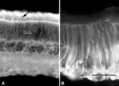
Figure 2. TAPA in the Injured Rat Retina
Sections of injured retina stained with the AMP1 antibody (A) demonstrate higher levels of immunoreactivity than seen in normal retina. A very distinct line of separation is apparent at the junction between the outer nuclear layer and the photoreceptor layer. High levels of immunoreactivity are apparent throughout the layers of the retina. Notice the fine AMP1 labeled projections extending through the outer segments of the photoreceptors (arrow). Sections of the injured retina stained for GFAP (B) show high levels of immunoreactivity in the ganglion cell layer as well as in the filaments extending throughout the layers of the retina. The cellular layers of the retina are indicated: ONL, outer nuclear layer; INL, inner nuclear layer; GCL, ganglion cell layer. The scale bar in B represents 20 µm.