![]() Figure 1 of
Ostrer, Mol Vis 4:28, 1998.
Figure 1 of
Ostrer, Mol Vis 4:28, 1998.
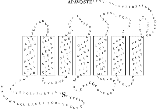
Figure 1. A secondary structure model of green cone opsin
The location of the N32S mutation is highlighted. The last eight amino acid residues shown in bold are the 1D4 epitope, which was added by in vitro mutagenesis.
