![]() Figure 8 of
Stenkamp, Mol Vis 4:26, 1998.
Figure 8 of
Stenkamp, Mol Vis 4:26, 1998.
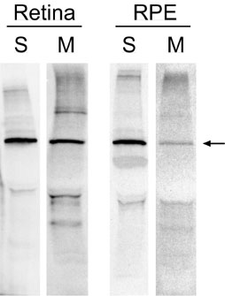
Figure 8. Immunoprecipitation of [35S]IRBP from zebrafish retina and RPE
The isolated retina and RPE-eyecup were incubated in the presence of [35S]methionine for 4 h after which IRBP was immunoprecipitated from the retina and RPE soluble fractions (S) and incubation media (M). Arrow indicates [35S]IRBP. Fluorograms: Lanes S, ~4 days exposure; Lanes M, ~7 days exposure.