![]() Figure 7 of
Stenkamp, Mol Vis 4:26, 1998.
Figure 7 of
Stenkamp, Mol Vis 4:26, 1998.
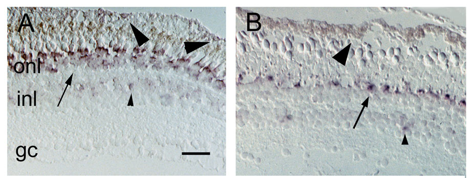
Figure 7. Expression of IRBP mRNA in cryosections from adult albino zebrafish retina and RPE
(A) Light-adapted retina, both rods and cones are labeled (arrow), as are scattered cells in the INL (small arrowhead). The basal region of the lightly-pigmented RPE is also labeled (large arrowheads); scale bar = 50 µm. (B) Dark-adapted retina, a subset of cones is labeled (arrow), as are some cells in the INL (small arrowhead). The dark-adapted RPE shows a distribution of pigmentation (large arrowhead) that may obscure labeling, making it difficult to discern whether this tissue is expressing IRBP mRNA.