![]() Figure 3 of
Stenkamp, Mol Vis 4:26, 1998.
Figure 3 of
Stenkamp, Mol Vis 4:26, 1998.
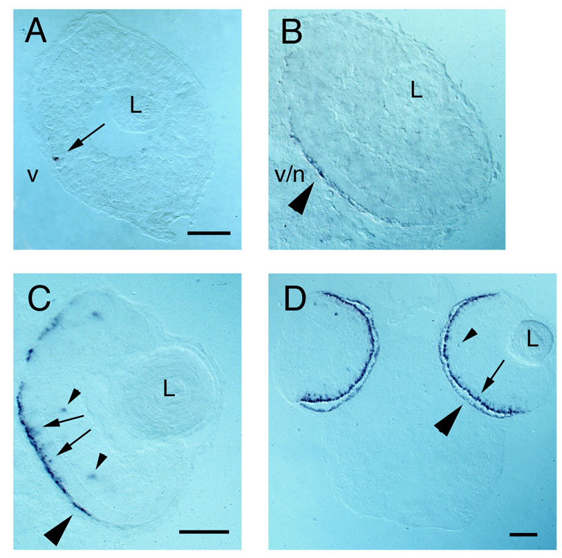
Figure 3. Expression of IRBP mRNA in embryonic zebrafish cryosections
All sections were hybridized with IRBP cRNA. (A) 51 hours post-fertilization (hpf) zebrafish section, arrow indicates a single labeled cell in the photoreceptor layer. The adjacent RPE is not labeled; v, ventral; L, lens. (B) 54 hpf zebrafish section, large arrowhead indicates region of RPE that is labeled; adjacent photoreceptors are not labeled; v/n, ventronasal. (C) 58 hpf zebrafish section, arrows indicate labeled differentiating photoreceptors. Large arrowhead indicates region of RPE that is labeled while adjacent photoreceptor cells are not, small arrowheads indicate labeled INL cells; L, lens. (D) 81 hpf zebrafish section, arrow indicates extensive labeling of photoreceptors, large arrowhead indicates extensive labeling of RPE, small arrowhead indicates labeled INL cell; L, lens. Scale bars = 50 µm; bar in A applies to B.