![]() Figure 1 of
Stenkamp, Mol Vis 4:26, 1998.
Figure 1 of
Stenkamp, Mol Vis 4:26, 1998.
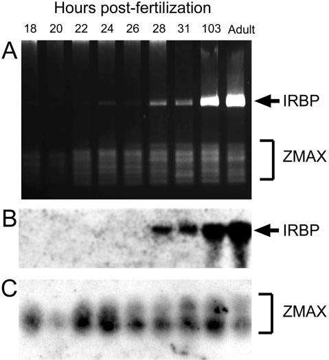
Figure 1. Temporal pattern of IRBP mRNA expression during zebrafish development
(A) RT-PCR amplification of a 530-bp IRBP mRNA fragment from whole zebrafish embryos at various times after fertilization (time indicated above each lane, 3% MetaPhor agarose (FMC) stained with ethidium bromide). IRBP mRNA (arrow) is first detectable at 24 to 28 hours post-fertilization (hpf). Zebrafish max mRNA (zmax) was co-amplified as an internal standard. (B) and (C): Southern blot analysis of the gel in panel A. IRBP (B) and zmax (C) RT-PCR fragments were probed with [[gamma]P]ATP-labeled oligonucleotides internal to those used for RT-PCR.