![]() Figure 5 of
Tao, Mol Vis 4:25, 1998.
Figure 5 of
Tao, Mol Vis 4:25, 1998.
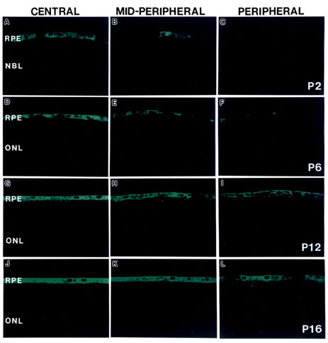
Figure 5. Expression of RGR in the developing mouse retina
Immunofluorescence was observed initially in RPE cells in the central retina at developmental stage P2 and increased thereafter from the central to the peripheral retina. The RPE cells during the embryonic period show no immunoreactivity (results not shown). The series of images encompasses the choroid, RPE and the outer portion of the neuroretina at P2 (A-C), P6 (D-F), P12 (G-I) and P16 (J-L). The neuroblastic layer (NBL) and the outer nuclear layer (ONL) are labeled.