![]() Figure 4 of
Bernstein, Mol Vis 1998;
4:24.
Figure 4 of
Bernstein, Mol Vis 1998;
4:24.
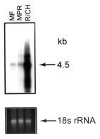
Figure 4. Northern analysis of total RNA using a cDNA probe from TIMP-3
Total RNA (5 mg) from each of pooled human fovea (HF); pooled human midperipheral retina (HPR); washed (RPE free) human choroidal tissue (Choroid); isolated, washed human RPE (RPE); pooled monkey fovea (MF); pooled monkey midperipheral retina (MPR); and monkey mixed choroid/RPE tissue (R/CH) were denatured, electrophoresed, transferred to nylon membranes and reacted with the radiolabelled cDNA probes as described in methods. Following hybridization, blots were stringently washed (63 °C/0.2X SSC), and exposed to X-ray film at -70 °C for varying lengths of time. Shown below each autoradiograph are the ethidium bromide stained 18S ribosomal RNA bands from the original agarose gel from which the analyzed blot was made. The ethidium bromide stained 18S rRNA bands were analyzed densitometrically and used as internal RNA loading standards. RDS to normalize variation in individual RNA loading [26].
Figure 4A. Analysis of expression from human fundus tissues, 14 day exposure
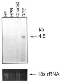
Figure 4B. TIMP-3 expression in rhesus monkey tissue, 5 day exposure