![]() Figure 1 of
Bernstein, Mol Vis 1998;
4:24.
Figure 1 of
Bernstein, Mol Vis 1998;
4:24.
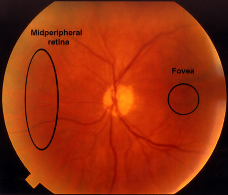
Figure 1. Human and rhesus monkey retinal regions used in the study
Demarcated areas indicate retinal regions used for the study. Human donor eyes were dissected on ice; the central 1.5 mm diameter (circle) surrounding the foveolar umbo, as well as a strip of midperipheral retina (oval area) were freed from underlying RPE and choroidal tissue and flash frozen on dry ice. Dissection of rhesus monkey retina was similar, except that the diameter of the dissected foveal tissue was 1.0 mm, due to the smaller size of the monkey eye. Pooled tissue from five individuals was used for total RNA isolation. Human RPE cells were isolated by washing the anterior surface of the choroid/RPE complex with iced phosphate buffered saline, centrifuged at low speed, and stored as a pellet prior to RNA extraction. Pooled RPE/choroid from monkey was flash frozen without further dissection. Total RNA was isolated was described in methods.