![]() Figure 2 of
Aguirre, Mol Vis 4:23, 1998.
Figure 2 of
Aguirre, Mol Vis 4:23, 1998.
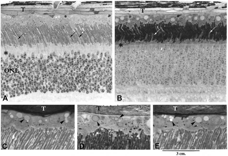
Figure 2. Retinal morphology in csnb
Histologic sections of plastic embedded retina from a 4.3-month-old dog affected with congenital stationary night blindness. Sections are taken from the tapetal zone (T) in the superior quadrant, near the posterior pole (A) or the mid-periphery (B). The retina is of normal thickness, and there is a normal number of photoreceptor cells and nuclei in the outer nuclear layer (ONL). The rod outer segments appear slightly irregular, particularly in the posterior pole (A), but are structurally better preserved in the mid-periphery (B). The variable shortening of rod inner segments results in outer segments of differing lengths. Cone inner segments appear elongated and distinct (A, B-oblique arrows). Cytoplasmic inclusions are present in the RPE (C-E, arrowheads). These occur as single small inclusions, or form aggregates or larger inclusions that can be homogeneous or vacuolated. * external limiting membrane. (A and B x500; C-D x1250).