![]() Figure 1 of
Aguirre, Mol Vis 4:23, 1998.
Figure 1 of
Aguirre, Mol Vis 4:23, 1998.
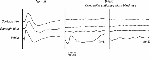
Figure 1. Electroretinographic responses in csnb
Representative dark adapted ERG responses recorded from a normal dog (left) and two briard dogs with csnb (center and right). The normal dog responds to scotopically balanced red and blue light stimuli with responses which are similar in waveform and amplitude. The response to a single high intensity (4.0 log-foot Lamberts) white light stimulus is biphasic, with a prominent a-wave, and an overall shorter latency b-wave response. In contrast, dogs affected with csnb have minute responses which are barely discernible over the baseline noise. When signal averaged over eight responses (n = 8), a distinct ERG response is evident, and the waveform is more characteristic of the normal response in the younger (4 months, center column) than in the older (2 years, right column) animal. This difference is not a characteristic finding with aging. (n = 8) is the number of responses averaged; vertical calibration is 170 µV for single responses, and 12 µV for averaged responses; horizontal calibration = 50 ms.
