![]() Figure 5 of
Kay, Mol Vis 4:22, 1998.
Figure 5 of
Kay, Mol Vis 4:22, 1998.
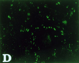
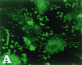
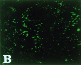
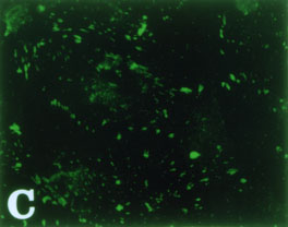
Figure 5. Immunofluorescent analysis of vinculin in CECs
Cells modulated with FGF-2 were treated with inhibitors in the presence of FGF-2 for 24 h. Cells were stained with anti-vinculin antibody, processed and analyzed on confocal microscopy as described in the text. (X 400)
(A) normal CECs
(B) cells treated with FGF-2
(C) cells treated with FGF-2 and LY294002 (20 µM)
(D) cells treated with FGF-2 and genistein (10 µM)