![]() Figure 2 of
Kay, Mol Vis 4:22, 1998.
Figure 2 of
Kay, Mol Vis 4:22, 1998.
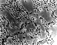
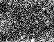
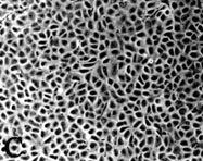
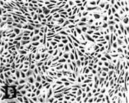
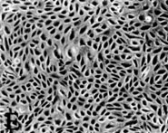
Figure 2. The effect of inhibitors on cell morphology of CECs modulated with FGF-2
The first passage CECs were treated with FGF-2 (10 ng/ml) supplemented with heparin (10 µg/ml) upon plating. On day 4, in which cellular morphology was changed to elongated fibroblastic shape, the cells were exposed to inhibitors for 48 h. (x150)
(A) normal CECs
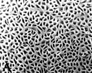
(B) CECs treated with FGF-2
(C) CECs treated with FGF-2 and 20 µM LY294002
(D) CECs treated with FGF-2 and 10 µM genistein
(E) CECs treated with FGF-2 and 100 nM wortmannin
(F) CECs treated with 40 µMLY294002