![]() Figure 10 of
Kay, Mol Vis 4:22, 1998.
Figure 10 of
Kay, Mol Vis 4:22, 1998.
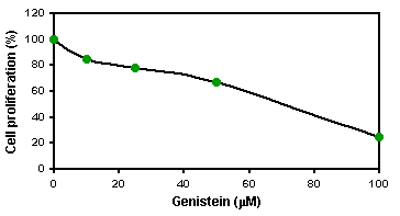
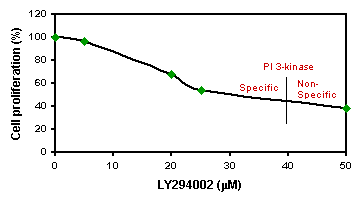
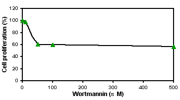
Figure 10. The effect of inhibitors on cell proliferation mediated by FGF-2 in CECs
The first passage CECs were plated into a 96-well plate and treated with one of the following conditions for 24 h when cells reached 80% confluency: FGF-2, FGF-2 and genistein in concentration ranging from 10 to 100 µM (top panel); FGF-2 and LY294002 in concentration ranging from 5 to 50 µM (middle panel); and wortmannin in concentration ranging from 10 to 500 nM (bottom panel). The cell proliferation assay was performed using CellTiter 96RAQueous One Solution Cell Proliferation Assay (Promega) as described in the text. Inhibitory activity of the inhibitors were presented as percent of FGF-2 mediated cell proliferation.


