![]() Figure 4 of
Rozsa, Mol Vis 4:20, 1998.
Figure 4 of
Rozsa, Mol Vis 4:20, 1998.
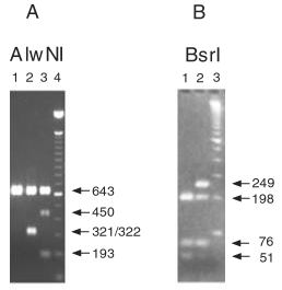
Figure 4. Heterozygous TIGR/MYOC sequence variants alter restriction sites
PCR-amplified fragments were digested with restriction enzymes as described in Materials and Methods. Arrows indicate fragment sizes on the right of each figure. A. Creation of new TIGR/MYOC exon 3 AlwNI sites in Fragment K. Lanes: 1. Unaffected control, 2. UM:JG1 proband (Pro370Leu), 3. UM:GL7 proband (Thr325Thr), 4. 100 base pair marker. B. Loss of TIGR/MYOC exon 3 BsrI site in Fragment L. Lanes 1. Unaffected control, 2. UM:GL57 proband (Glu323Lys), 3. 100 base pair marker.