![]() Figure 2 of
Rozsa, Mol Vis 4:20, 1998.
Figure 2 of
Rozsa, Mol Vis 4:20, 1998.
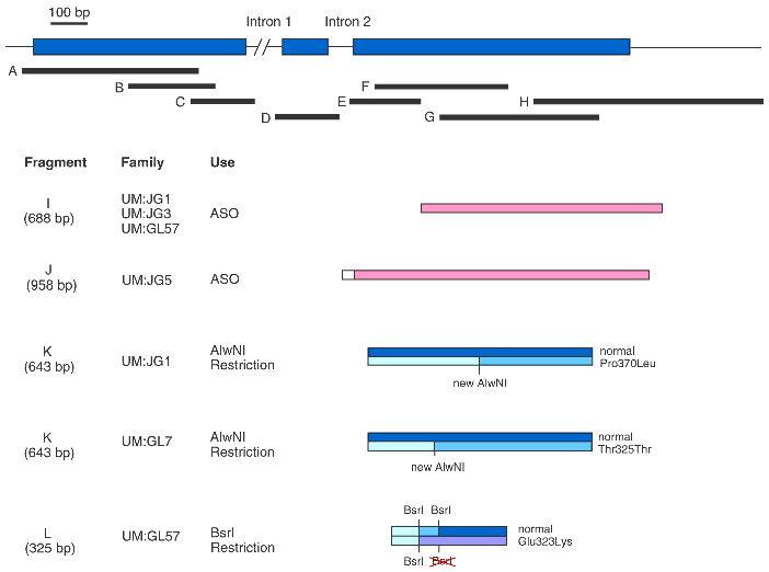
Figure 2. PCR-amplified TIGR/MYOC fragments used for sequencing, ASO, and restriction analysis
Gene structure of TIGR/MYOC is shown below with exons 1-3 in blue boxes. Fragments A-H (black boxes) were amplified by PCR using primers described in Table 1 and used as templates in subsequent DNA sequencing reactions. Fragments used for ASO (I, J) are shown by pink boxes. Fragments used for restriction analysis (K, L) to test for mutations in each family are shown as shaded blue boxes. All primers correspond to Table 1.