![]() Figure 6 of
El-Kabbani, Mol Vis 1998;
4:19.
Figure 6 of
El-Kabbani, Mol Vis 1998;
4:19.
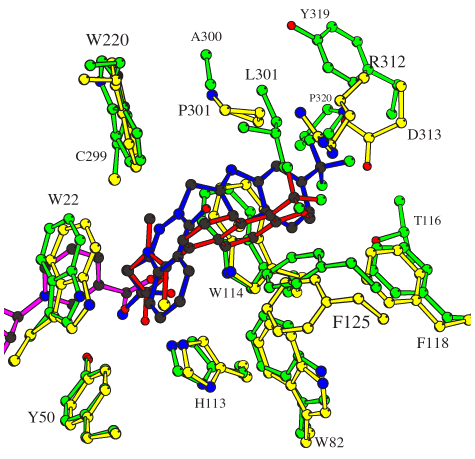
Figure 6. The inhibitor-binding site
Superimposed inhibitor-binding residues for ALR1 with bound tolrestat (amino-acid side-chains in yellow, inhibitor in red) and ALR2 with bound zopolrestat (amino-acid side-chains in green, inhibitor in blue). The inhibitor atoms are colored by type to illustrate potential hydrogen bonds (O in red, N in blue, S in yellow, F in green, C in black). For clarity, only the coenzyme molecule bound to ALR1 is shown in pink. Residues are labeled with residue type and number. The sequence numbering for aldehyde reductase (ALR1) is used [11].