![]() Figure 4 of
El-Kabbani, Mol Vis 1998;
4:19.
Figure 4 of
El-Kabbani, Mol Vis 1998;
4:19.
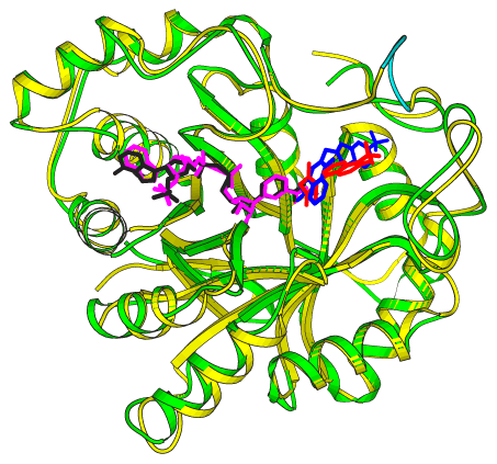
Figure 4. Backbone foldings of ALR1 and ALR2
Superposition of porcine ALR1-tolrestat (back bone in yellow, coenzyme in pink, inhibitor in red) and human ALR2-zopolrestat (back bone in green, coenzyme in black, inhibitor in blue) structures. The eight residue insertion segment in the C-terminal loop of ALR1 (residues 306-313) is shown in cyan. Ribbon drawings were prepared using MOLSCRIPT [37].
