![]() Figure 1 of
Tucker, Mol Vis 4:18, 1998.
Figure 1 of
Tucker, Mol Vis 4:18, 1998.
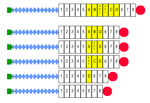
Figure 1. Tenascin-C Variants
Stick diagrams illustrating the domain organization of avian tenascin-C and a few of the known alternatively spliced variants. The EGF-like repeats are indicated with blue diamonds, the constant fibronectin type III repeats are numbered white boxes, the variable fibronectin type III repeats are lettered yellow boxes, and the fibrinogen-like terminal domain is a red circle. The tenascin-C shown at the top is hypothetical; it shows the relationships of the 6 variable repeats within the variable domain. At the bottom are 5 known splice variants (see [13] and [17] for other variants). Three contain 3 variable repeats and would have apparent molecular weights of approximately 230 kDa (Tn230). One has a single repeat and would have an apparent molecular weight of 200 kDa (Tn200). The tenascin-C at the bottom does not have any variable repeats, and would have a molecular weight of 190 kDa (Tn190).