![]() Figure 3 of
Howell, Mol Vis 4:15, 1998.
Figure 3 of
Howell, Mol Vis 4:15, 1998.
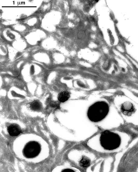
Figure 3. Immunoelectron micrograph of human ciliary body stained with rabbit antiserum against carboxy-terminal peptide of human placenta amine oxidase
Sections of human ciliary body produced by ultracryomicrotomy were stained with rabbit antiserum against the carboxy-terminal peptide of human placenta amine oxidase and a colloidal-gold-conjugated secondary antibody. Numerous colloidal gold particles (black dots) are localized to the plasma membrane of an inner epithelial cell (top half of micrograph), visible in may areas as a thin, lucent line. A small number of gold particles are localized to the surface of an adjacent outer epithelial cell (bottom half of micrograph) and to cytoplasm of both cells. The latter cell contains numerous electron-dense pigment granules; no significant staining of the granules is present. The clear halos surrounding the granules and the small, bubble-like lucencies within some of them are most likely artifacts of processing and embedding of the sections.