![]() Figure 1 of
Howell, Mol Vis 4:15, 1998.
Figure 1 of
Howell, Mol Vis 4:15, 1998.
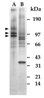
Figure 1. SDS-PAGE of human ciliary body proteins isolated by affinity chromatography with 2B4.14.1
Samples were reduced, mock-digested (lane A) or digested with a mixture of endoglycosidase F/N-glycosidase F (lane B), and separated on a 5-15% gradient gel, which was then silver stained. The major bands at 80, 100, and 130 kDa (arrowheads) show a downward shift in apparent molecular weight following glycosidase digestion. Each lane contains 2B4.14.1 ligands isolated from approximately 200 µg of whole ciliary body protein. The dark band at approximately 35 kDa in lane B is the added glycosidase.