![]() Figure 3 of
Zhu, Mol Vis 1998;
4:13.
Figure 3 of
Zhu, Mol Vis 1998;
4:13.
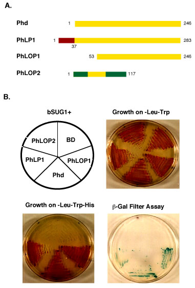
Figure 3. Interaction of Phd isoforms with bSUG1 in yeast.
(A) Schematic alignment of the domains representing the amino acid sequences of each Phd isoform which was subcloned into the GAL4 BD (not shown in the figure) at their amino-terminus. The identical sequences are indicated with yellow bars. The unique domains for either PhLP1 or PhLOP2 are shown in different colors. (B) Assays of HIS3 and LacZ reporter gene expression. The yeast reporter strain CG-1945 was cotransformed with AD-bSUG1 (clone7) and each of the BD-Phd isoforms (top left panel). A pool of colonies from -Leu-Trp plates was streaked onto selective medium with histidine (-Leu-Trp) (top right panel) or without histidine (-Leu-Trp-His) (lower left panel). The plates were incubated at 30 °C for 4 days. Cells grown on -Leu-Trp-His plates were transferred onto a Whatman #1 filter, lysed by rapid freezing in liquid nitrogen and thawing at room temperature. The filter was then placed on top of another a Whatman #1 filter pre-soaked in Z buffer with X-gal and incubated at 30 °C. The positive blue color reaction developed within 2 hours (lower right panel).