![]() Figure 5 of
Townes-Anderson, Mol Vis 4:12, 1998.
Figure 5 of
Townes-Anderson, Mol Vis 4:12, 1998.
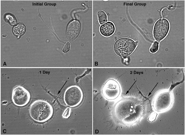
Figure 5. Cell contacts form after optical trapping
Cell groups which were formed by optical tweezers micromanipulation were followed over time to observe the initial stages of contact formation. Here, the initial group contained a cone cell (left) and a multipolar neuron (right). A rod cell was placed in this group. Prominent growth is exhibited by the moved rod cell. A large process is directed toward the multipolar neuron. Arrows, first contacts at 1 and 2 days in vitro.