![]() Figure 4 of
Townes-Anderson, Mol Vis 4:12, 1998.
Figure 4 of
Townes-Anderson, Mol Vis 4:12, 1998.
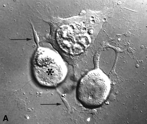
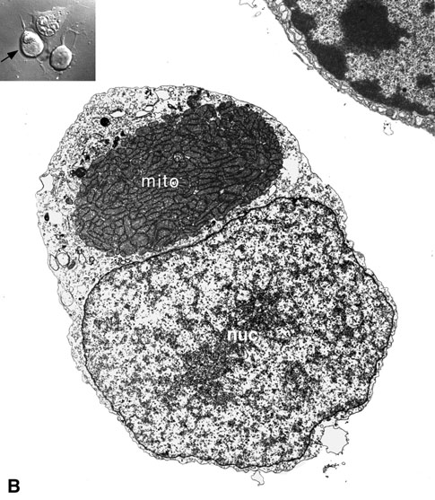
Figure 4. Fine structure of trapped cells is normal
(A) An optically trapped and repositioned cone cell (*) showing process outgrowth and formation of synaptic-like varicosities (arrows) after 2 days in vitro.
(B) Electron microscopy demonstrated that the cell soma contained the characteristic complement of cell organelles. Note the normal appearance of mitochondria (mito) and the nucleus (nuc). Inset, arrow indicates location of the electron micrograph.
(C) Presynaptic varicosities were filled with synaptic vesicles (sv). A multivesicular body is also present in this varicosity. Inset, arrow indicates location of the electron micrograph.

(D) Synaptic ribbons (rb, arrows) were present in the cell body. Presumably, they moved from the axon into the cell soma after cell isolation and as the cell began to regenerate new neuritic processes. They remain surrounded by a halo of synaptic vesicles.
