![]() Figure 2 of
Townes-Anderson, Mol Vis 4:12, 1998.
Figure 2 of
Townes-Anderson, Mol Vis 4:12, 1998.
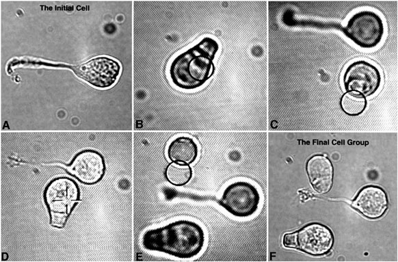
Figure 2. Formation of cell groups
Using a sealed culture dish, an adherent cell was chosen from the antibody-coated side of the dish; nonadherent cells were selected from the opposite side, and placed next to the chosen cell. Here, the adherent cell was a multipolar neuron (A); then a nonadherent rod cell (B-D) and a cone cell (E-F) were optically trapped and moved to it. Circles indicate the approximate location of the optical trap; cross indicates laser off. A and F, images obtained with CCD camera mounted on the microscope; B-E, images obtained from video tape of monitor as cells were being moved.