![]() Figure 1 of
Townes-Anderson, Mol Vis 4:12, 1998.
Figure 1 of
Townes-Anderson, Mol Vis 4:12, 1998.
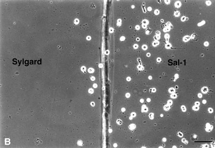
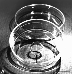
Figure 1. Culture dish preparation
(A) A well was created in a 35 mm plastic dish by drilling a hole and affixing a glass coverslip to the dish [11]. One side of the well was coated with an adherent substrate consisting of a monoclonal antibody, Sal-1 [12]; the other side was made less adherent by applying a thin coat of the elastomer Sylgard (Dow Corning Corp.). The juncture of the two surfaces was marked with an etched line on the back surface of the glass coverslip (arrow). Bar = 1 cm.
(B) After 8 days in culture the difference in cell adhesion was apparent with growing cells attached mainly on the surface coated with Sal-1. Bar = 100 µm.