![]() Figure 2 of
Xu, Mol Vis 4:10, 1998.
Figure 2 of
Xu, Mol Vis 4:10, 1998.
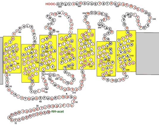
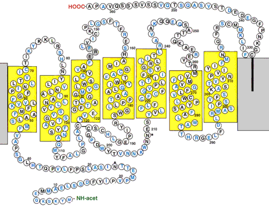
Figure 2. Predicted secondary structures of the red and blue cone pigments
The transmembrane domains (boxed) were defined based on Kyte-Doolittle hydropathy plots and the comparison with bovine rhodopsin. The Lys residues at chromophore attachment sites and the Schiff's base counterion Glu residues are indicated by diamond boxes and numbering their positions. The ERY motif is shaded. The potential glycosylation sites are indicated by an asterisk (*). Residues identical to the salamander rhodopsin are in black.
Figure 2A. Predicted secondary structure of the red cone pigment

Figure 2B. Predicted secondary structure of the blue cone pigment
