![]() Figure 5 of
Chitpinityol, Mol Vis 4:1, 1998.
Figure 5 of
Chitpinityol, Mol Vis 4:1, 1998.
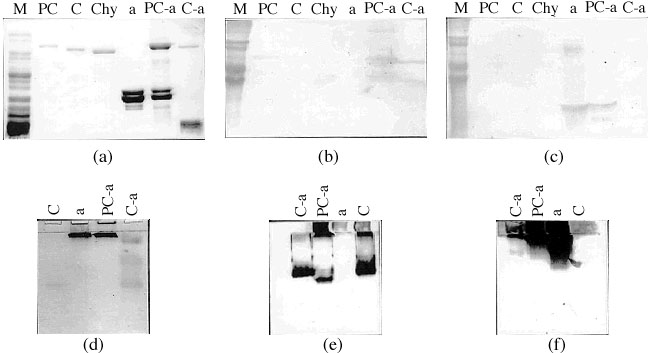
Figure 5. Refolding of wild-type prochymosin in the presence of [alpha]-crystallin.
Coomassie blue stained SDS-PAGE and Western blot analysis of the refolding of wild-type prochymosin in the presence of [alpha]-crystallin. The experiment of panel (a) is identical to that of (b) and (c) whereas (d) is identical to (e) and (f). (a): 10% SDS-PAGE. (d): 10% nondenaturing gel. (b), (e): Western blots probed with polyclonal antichymosin antibodies. (c), (f): Western blots probed with polyclonal anti-[alpha]-crystallin antibodies. The lanes are: molecular weight marker ("M"), authentic chymosin ("Chy"), [alpha]-crystallin ("a"), prochymosin ("PC"), chymosin ("C"), prochymosin refolded with [alpha]-crystallin ("PC-a"), and chymosin from prochymosin folded with [alpha]-crystallin ("C-a").