Figure 5. Pedigree diagram and phenotypic photographs. A-B: Pedigree of the families with sequencing of pathogenic variants in KERA. C-F: Slit-lamp photographs and photograph of diameter of cornea of patients with pathogenic variants of KERA identified in our cohort.
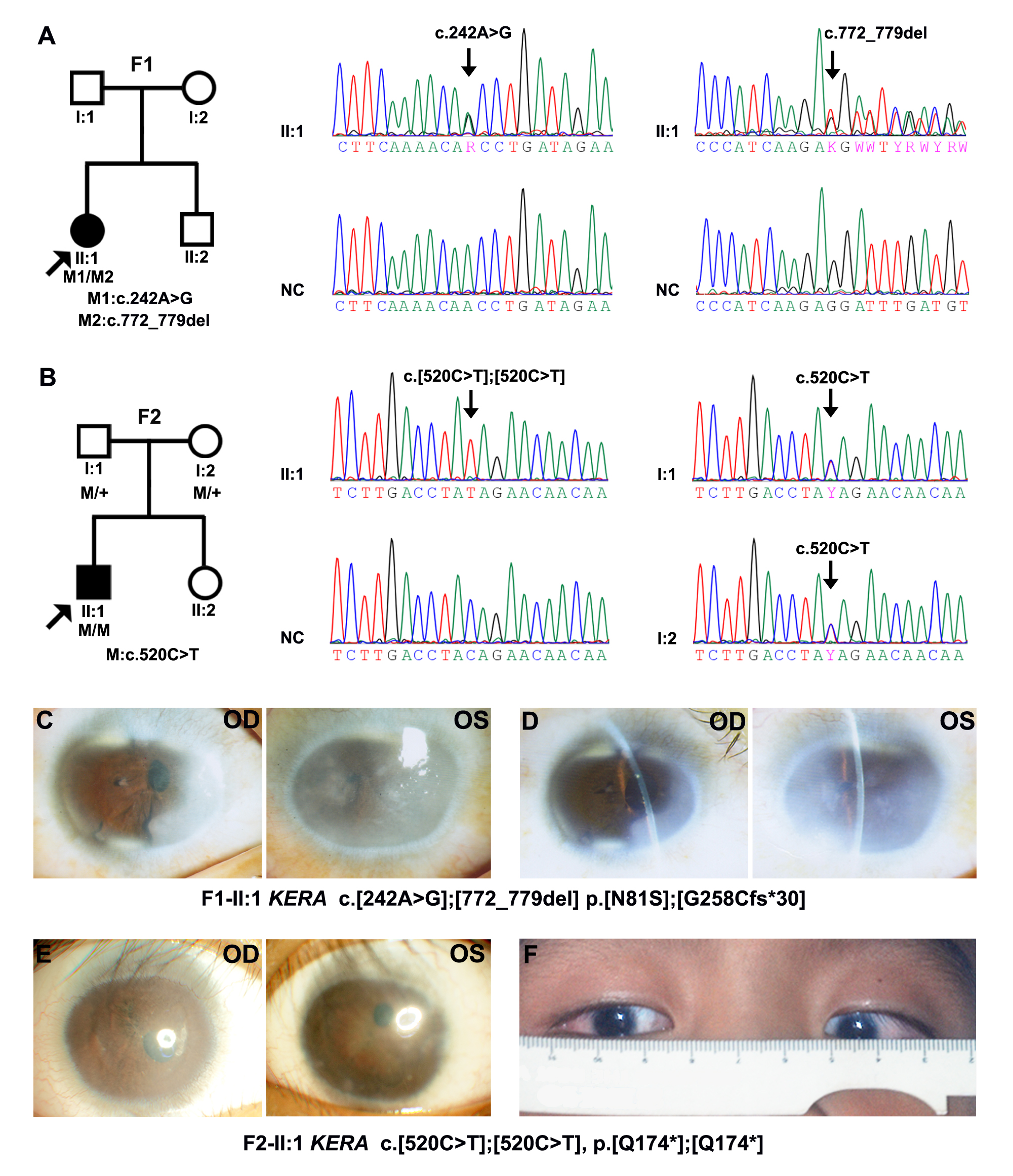
 Figure 5 of
Zhu, Mol Vis 2026; 32:70-82.
Figure 5 of
Zhu, Mol Vis 2026; 32:70-82.