Figure 1. Retinal imaging of V1 family: Composite montage of color fundus photographs (upper panels) and corresponding optical coherence
tomography (OCT) B-scans (lower panels) of 24 affected members from the original 765 V1 pedigree. Images are aligned by subject,
with right and left eyes shown side-by-side within each panel.
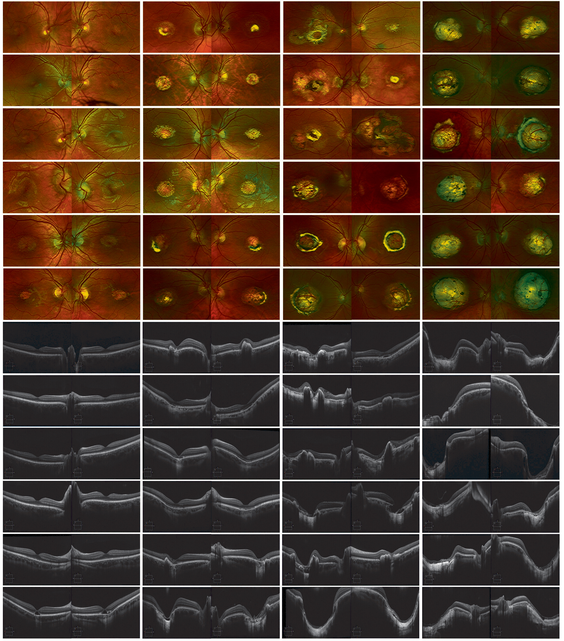
 Figure 1 of
Small, Mol Vis 2026; 32:451-463.