Figure 3. Effects of PEDF[H105A] protein on CRX in mouse photoreceptors. A: Treatment scheme illustrating wild-type mouse retinal explants treated without or with 20 nM PEDF[H105A] for 48 h. B: Representative regions of interest (ROIs) from confocal microscopy images of the retinal explants with CRX immunofluorescence
staining (green) and DAPI staining of photoreceptor nuclei (pseudo-gray). Data are representative of three biological replicates
(three eyes) and four technical replicates (ROIs per eye). Mice were 3 months old. C: CRX mean fluorescence intensity (MFI) was measured within each ROI and normalized to the number of DAPI-positive nuclei;
each point represents one explant from one of three independent mice (n = 3). Data are shown as mean ± SD. Statistical significance
was determined by an unpaired t test: *p < 0.05.
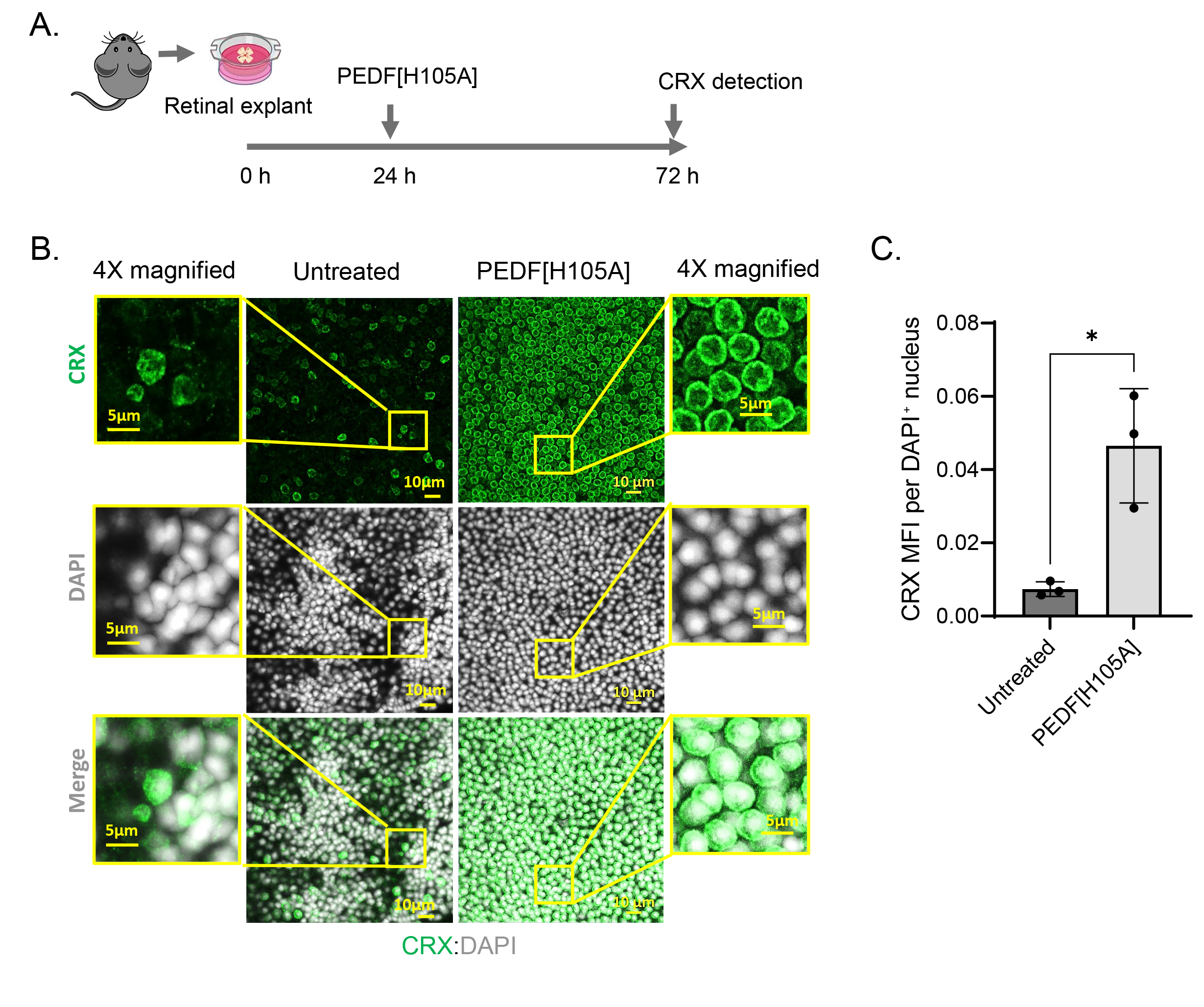
 Figure 3 of
Dixit, Mol Vis 2026; 32:435-449.