Figure 2. Reduced PDE6A immunoreactivity and pan-histone acetylation in photoreceptors of Serpinf1−/− mice. A: Representative regions of interest (ROIs) from Serpinf1+/+ and Serpinf1−/− retinas showing PDE6A immunoreactivity (red), pan-histone acetylation (green), and Hoechst nuclear staining (blue) by confocal
microscopy. Regions indicated by yellow squares are shown at higher magnification. B: Quantification of PDE6A mean fluorescence intensity (MFI). C: Quantification of pan-histone acetylation MFI. Quantification was performed using two ROIs from of three biological replicates.
Mice were 3 months old. *p < 0.05, **p < 0.01.
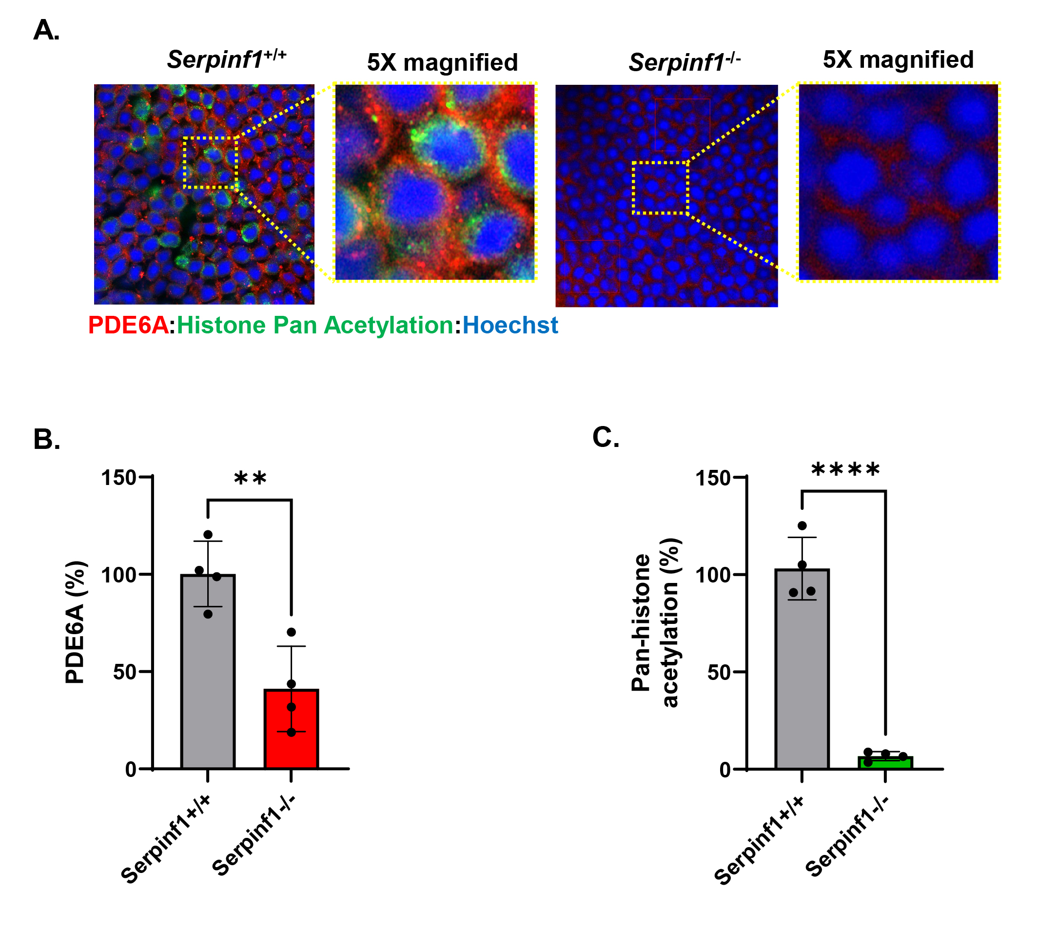
 Figure 2 of
Dixit, Mol Vis 2026; 32:435-449.