Figure 5. Schematic representation and three-dimensional structure of the keratocan protein. A: This panel shows the structural organization of keratocan, which consists of one leucine-rich repeat N-terminal (LRRNT)
domain and 11 leucine-rich repeat (LRR) domains composed of alternating α-helices and β-sheets. The positions of the six identified
KERA variants are indicated according to their amino acid locations within the protein domains. B: This panel shows the three-dimensional structures of the wild-type KERA protein and the mutant p.Asn176Lys and p.Ser270Leu
proteins. The mutant proteins exhibit minimal global structural deviations from the wild-type protein, with root mean square
deviation values of 0.188 Å and 0.204 Å, respectively, indicating preservation of the overall protein fold.
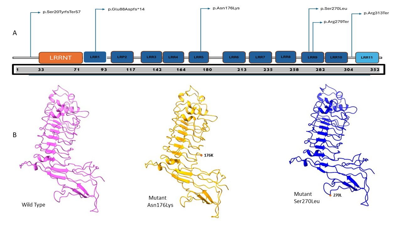
 Figure 5 of
Verma, Mol Vis 2026; 32:410-419.