Figure 3. Representative clinical images of the right eye of CP5a. A: Slit-lamp photograph shows diffuse stromal haze with a ground-glass appearance, markedly flattened corneal curvature, and
indistinct limbal architecture. B: Pachymetry map demonstrates an abnormal corneal thickness profile with relative central thickening and peripheral thinning.
C: Anterior segment optical coherence tomography (AS-OCT) image shows reduced corneal curvature and central corneal thickness
of approximately 450 µm, a thinner peripheral cornea, and a shallow anterior chamber. Fine iris details are limited, but imaging
suggests anterior iris insertion toward the posterior corneal surface. D: Specular microscopy image shows preserved endothelial cell morphology without significant pleomorphism or polymegathism.
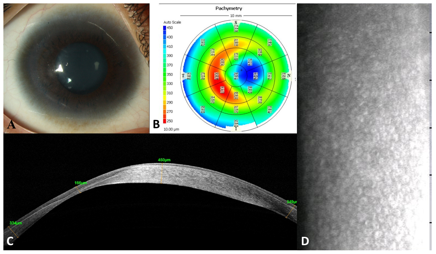
 Figure 3 of
Verma, Mol Vis 2026; 32:410-419.