Figure 2. Representative clinical images of CP1 in each eye. A, B: Slit-lamp photographs show markedly flattened corneal curvature and diffuse stromal haze with a ground-glass opacity. The
limbal architecture appears indistinct, blending with the sclera. The pupil is poorly visualized due to stromal opacity. C, D: Anterior segment optical coherence tomography (AS-OCT) cross-sectional images reveal marked corneal flattening with reduced
anterior curvature. The cornea appears thickened centrally relative to the periphery. There is a significantly shallow anterior
chamber, with anteriorly displaced iris tissue and iridocorneal adhesions, indicating abnormal anterior segment configuration.
The angle appears compromised, predisposing to angle-closure mechanisms. Stromal reflectivity is increased, corresponding
to corneal haze.
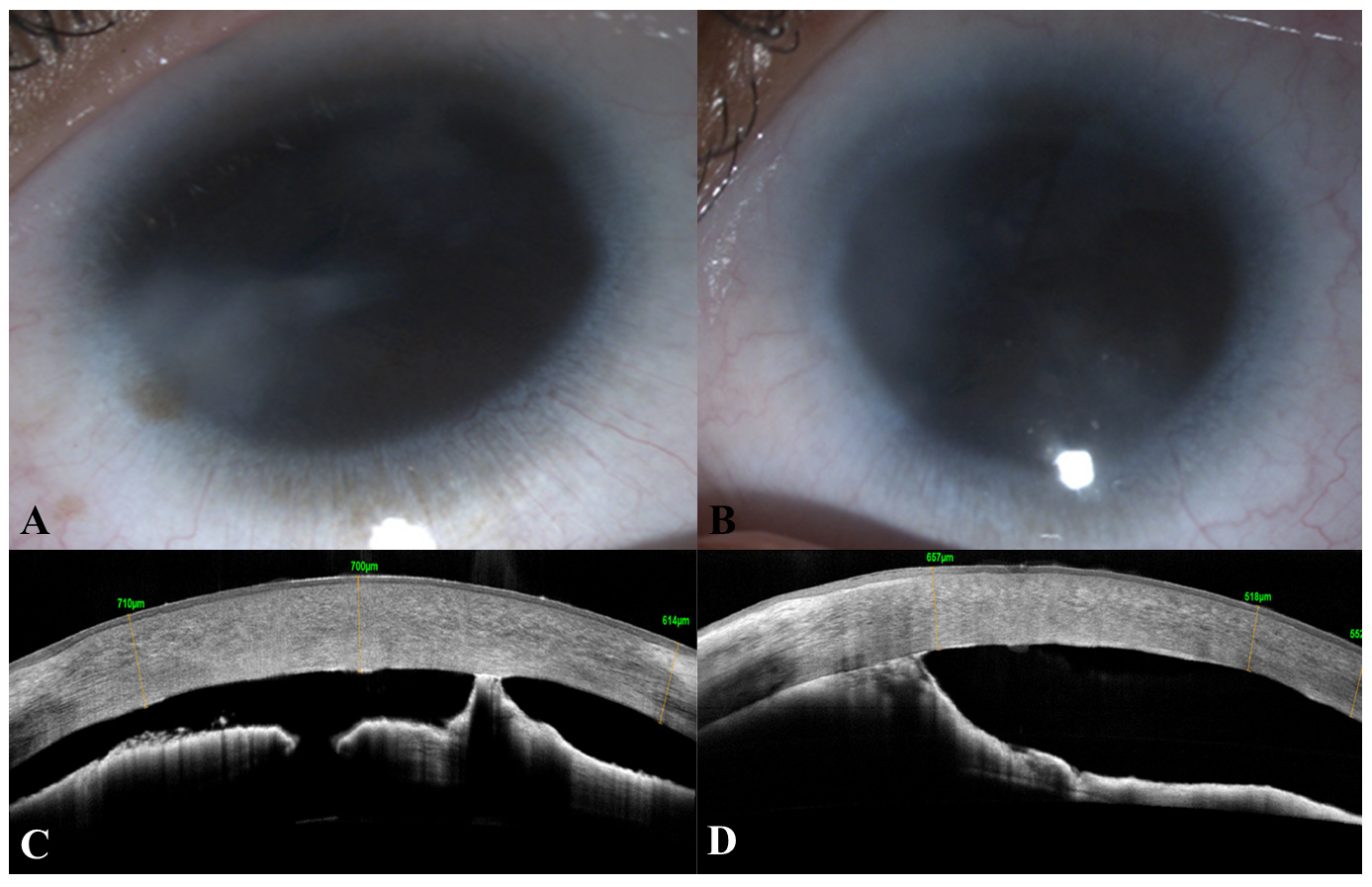
 Figure 2 of
Verma, Mol Vis 2026; 32:410-419.