Figure 7. HXTL ameliorates high glucose-induced dysregulation of the MEG3/miR-494–5p axis and PI3K/AKT/mTOR signaling, and alleviates
functional impairment in mRMECs. A: Dual-luciferase reporter assay confirming the direct targeting of MEG3 by miR-494–5p, n=3; B, C: Expression levels of lncRNA MEG3 and miR-494–5p in mRMECs under various treatments, n=3; D-G: Protein expression levels of p-PI3K, p-AKT, and p-mTOR with corresponding quantitative analysis, n=3; H: Assessments of cell viability, n=3; I-J: Assessments of proliferation and quantification of EdU-positive cells, n=3; K-M: Representative images and quantitative analysis of branch points and tube length from the tube formation assay, n=3; N: Endothelial permeability measured by FITC-dextran transendothelial flux, n=3. #p<0.05, ##p<0.01 versus normal group, @p<0.05, @@p<0.01 versus HG group; $p<0.05, $$p<0.01 versus HXTL group. mRMECs: mouse retinal microvascular endothelial cells, Edu: 5-ethynyl-2′-deoxyuridine, FITC: fluorescein
isothiocyanate.
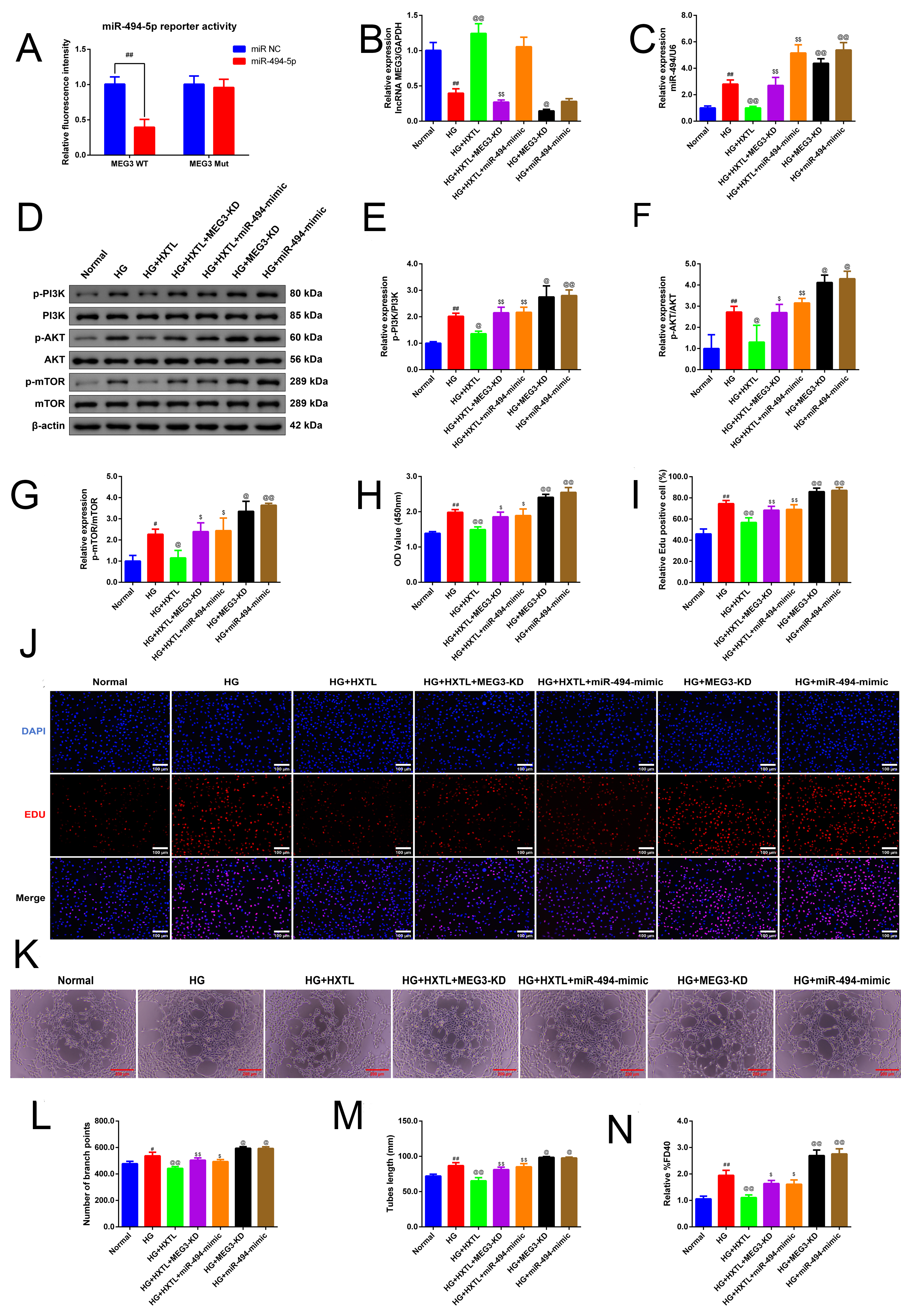
 Figure 7 of
Li, Mol Vis 2026; 32:370-386.