Figure 4. Effects of HXTL on apoptosis, VEGF expression, and inflammatory factors in DR mice. A, B: Representative images (A) and quantification (B) of TUNEL-positive apoptotic cells in retinal tissues (magnification × 200, scale bar: 50 μm; magnification × 400, scale
bar: 25 μm), n=3; F-G: ELISA was used for testing VEGF level (c), IL-1β (D) and Hcy (E) in serum and IL-1β (f), TNF-α (G) in retinal tissues of mice in each group, n=6; H-J: qRT-PCR was used for testing IL-1β, IL-6, TNF-α levels of the mice in each group, n=3; ▲▲p<0.01 versus control group, ★p<0.05, ★★p<0.01 versus DR group. TUNEL: TdT-mediated dUTP nick-end labeling, VEGF: vascular endothelial growth factor, IL-1β: interleukin-1
beta, Hcy: homocysteine, TNF-α: tumor necrosis factor-α.
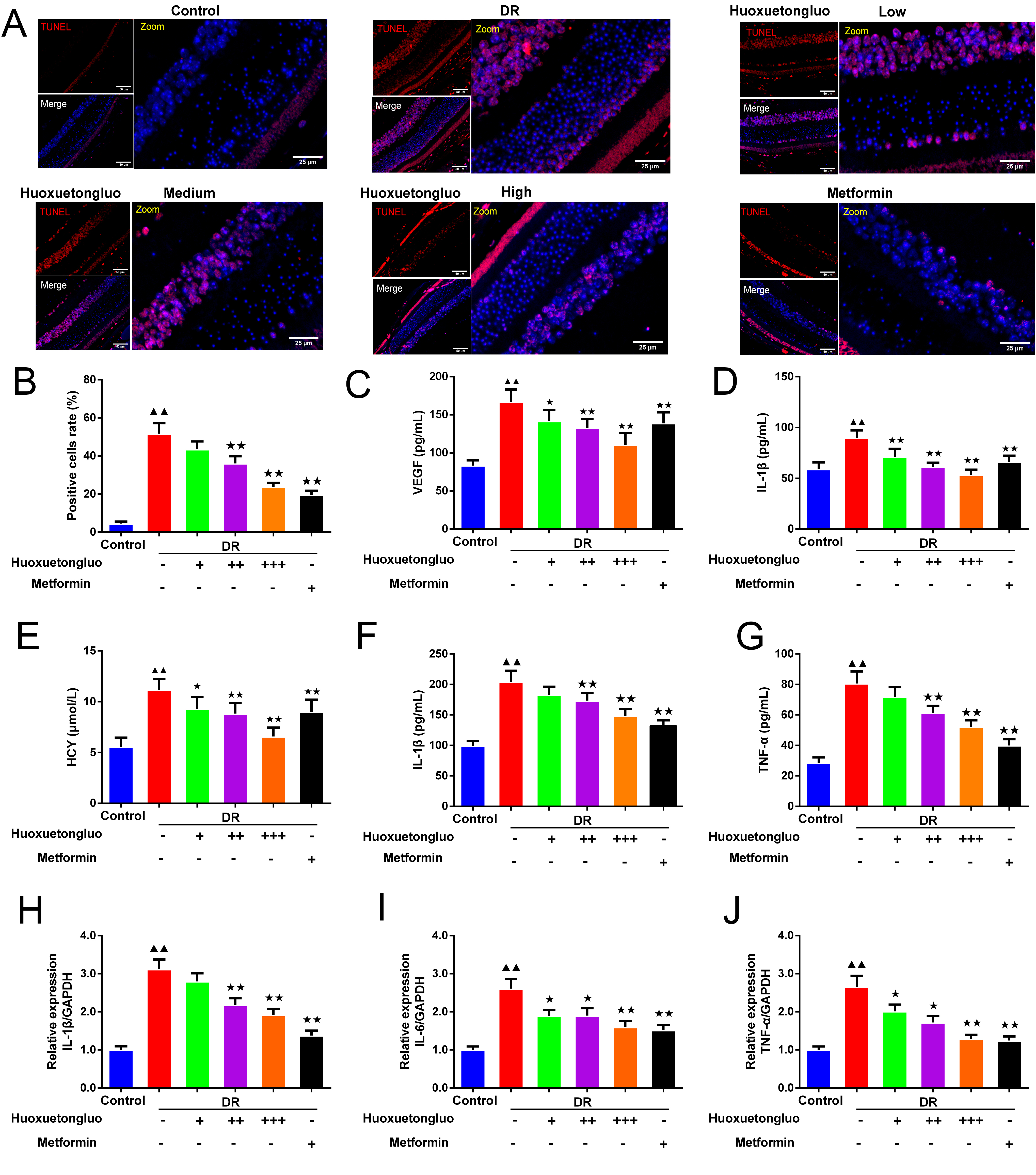
 Figure 4 of
Li, Mol Vis 2026; 32:370-386.