Figure 3. The effect of HXTL on histopathological damage and angiogenesis of DR in mice. A: The histomorphology of diabetic retinal tissues in mice was observed by HE staining (magnification × 200, scale bar: 100
μm; magnification × 400, scale bar: 50 μm); B-D: The positive expressions of CD31 and VEGFR of diabetic retinal tissues in mice were examined by IHC (magnification × 200,
scale bar: 100 μm; magnification × 400, scale bar: 50 μm). VEGFR: vascular endothelial growth factor receptor.
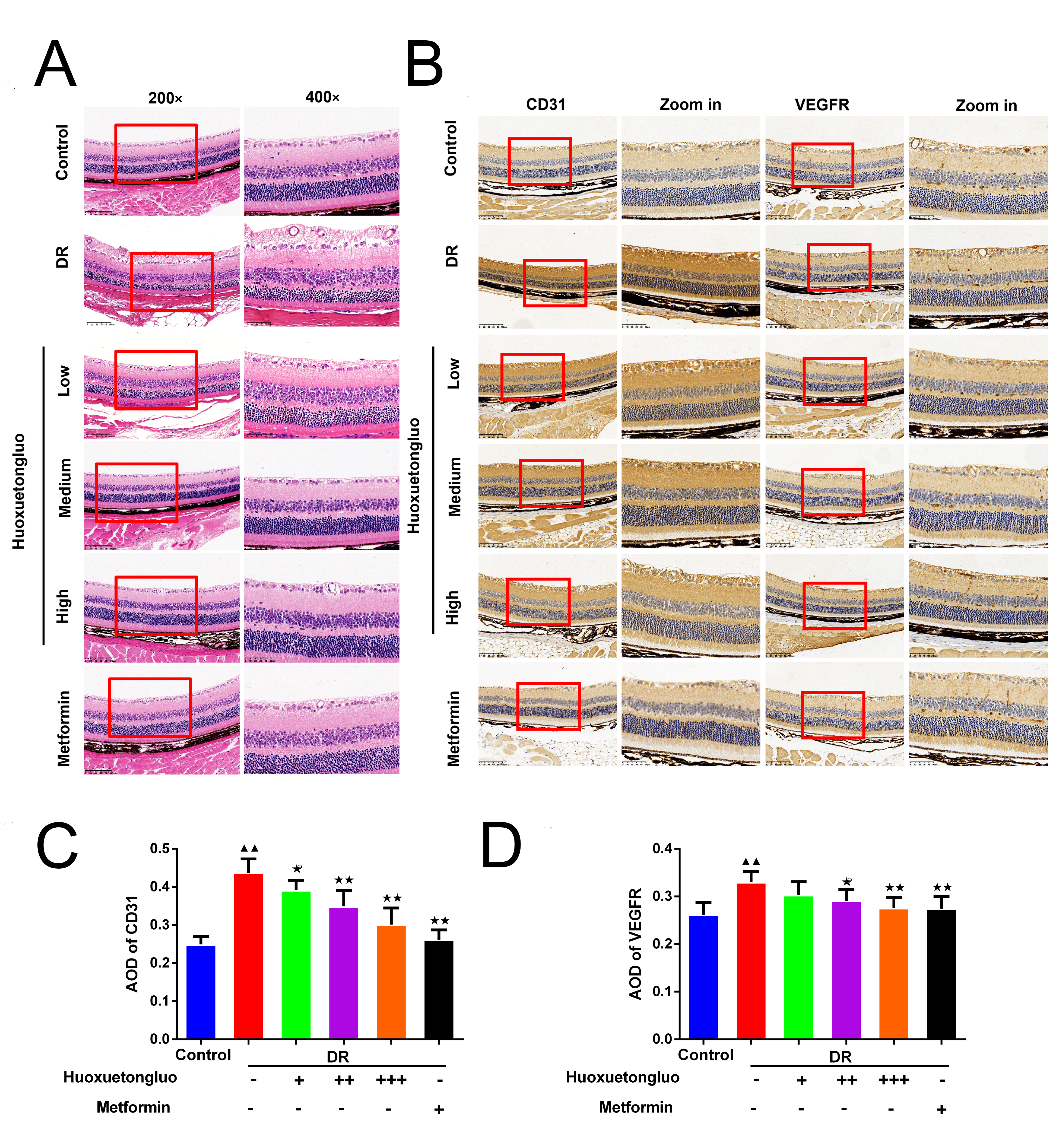
 Figure 3 of
Li, Mol Vis 2026; 32:370-386.