Figure 1. Aqp5 deficiency induces ferroptosis in lens capsules and lens epithelial cells (LECs) of mice. A: Transmission electron microscopy images reveal obvious ultrastructural alterations in LECs. B: BODIPY581/591 C11 staining demonstrates that the levels of lipid reactive oxygen species are significantly elevated in primary LECs isolated
from Aqp5−/− mice compared with primary LECs from Aqp5+/+ wild-type mice. C: Quantitative statistical analysis was conducted for the green-to-red fluorescence ratio obtained from BODIPY 581/591 C11 staining (n = 6). D: FerroOrange immunofluorescence staining shows that Fe2+ accumulation is markedly increased in primary Aqp5−/− LECs. E: Quantitative analysis was performed to measure the fluorescence intensity of FerroOrange probes (n = 6). F: Western blot bands display the protein expression levels of Nrf2, SLC7A11, SLC3A2, and GPX4 in LECs harvested from Aqp5+/+ and Aqp5−/− mice. G: Relative quantitative results of target protein expression were analyzed by ImageJ software. All quantitative data in this
figure are expressed as mean ± SD.
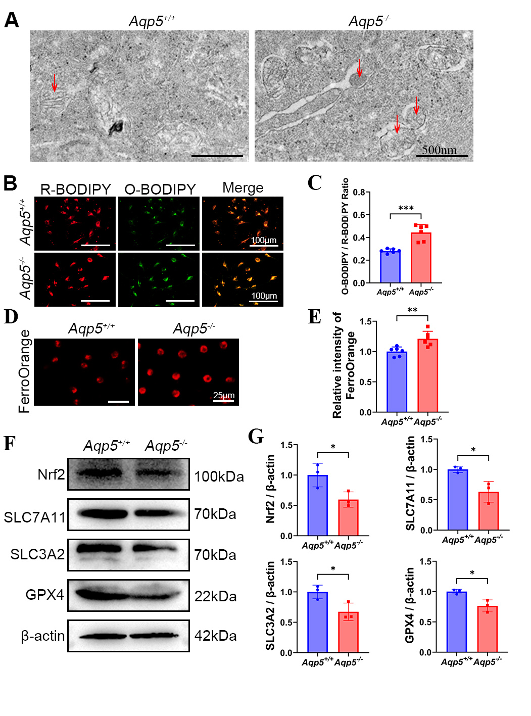
 Figure 1 of
Song, Mol Vis 2026; 32:354-369.