Figure 1. Representative SS-OCT image of the macula depicts a vertical cross-sectional image of the macula taken using swept-source
optical coherence tomography (SS-OCT). A. Choroidal thickness is measured from Bruch’s membrane to the choroid–scleral interface. B. The retinal layers are displayed from the internal limiting membrane to the outer segment–retinal pigment epithelium interface
of the photoreceptor layer.
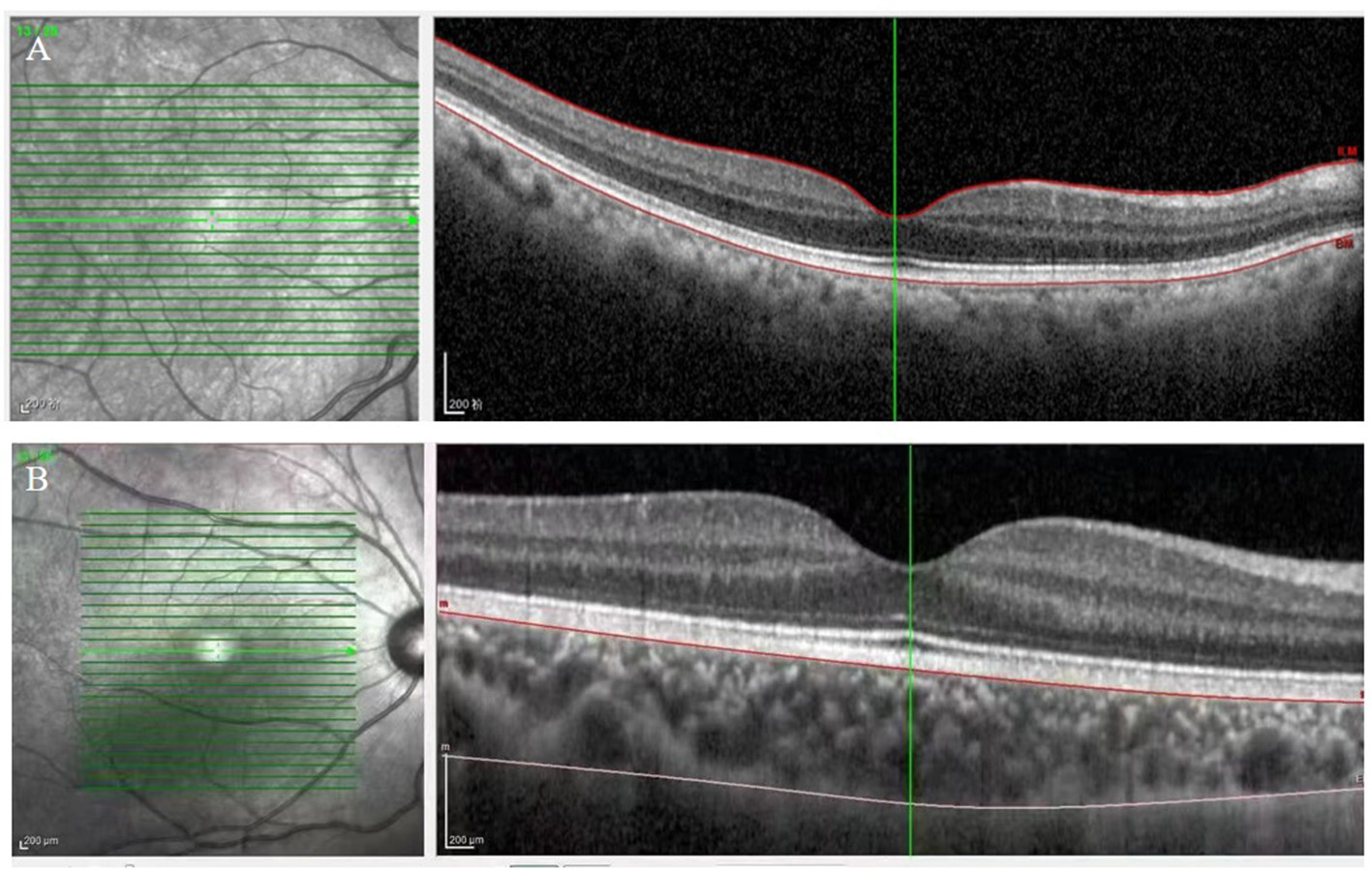
 Figure 1 of
Zhao, Mol Vis 2026; 32:343-352.