Figure 5. Expression of proinflammatory gene transcripts in human retinal pigment epithelial cells. After transfection with Zaire ebolavirus viral protein 24 (VP24) or no-insert control (NI) expression plasmid for 48 h, followed by polyinosinic-polycytidylic acid
(poly I:C) for an additional 4 or 24 h, cellular levels of levels of six proinflammatory gene transcripts were measured by
quantitative polymerase chain reaction and normalized to two stable reference genes (glyceraldehyde 3-phosphate dehydrogenase
[GAPDH] and peptidylprolyl isomerase A [PPIA]). An additional control replacing poly I:C with water was included for the 4-h
condition. In All Isolate graphs, circles represent mean expression in individual isolates, crossbars indicate mean, and error bars indicate standard
deviation (n = 4 isolates/condition). In Isolate 1–4 graphs, bars indicate mean normalized expression, and error bars indicate standard deviation (n = 4 replicates/condition).
CCL2, C-C motif chemokine ligand 2; ICAM1, intercellular adhesion molecule 1; IL1B, interleukin 1 beta; IL6, interleukin 6;
ND, transcript not detectable in ≥2 of 4 replicates; ns, not significant; TNF, tumor necrosis factor; VCAM1, vascular cell
adhesion molecule 1. *p < 0.05, **p < 0.01, ***p < 0.001, ****p < 0.0001.
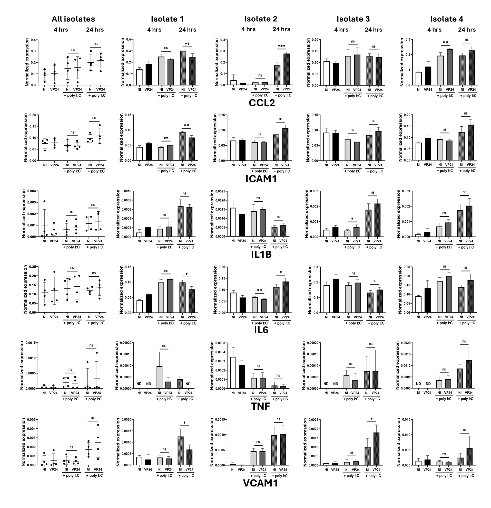
 Figure 5 of
Ashander, Mol Vis 2026; 32:34-48.
Figure 5 of
Ashander, Mol Vis 2026; 32:34-48.