Figure 3. Expression of antiviral gene transcripts in human retinal pigment epithelial cells. After transfection with Zaire ebolavirus viral protein 24 (VP24) or no-insert control (NI) expression plasmid for 48 h, followed by polyinosinic-polycytidylic acid
(poly I:C) for an additional 4 or 24 h, cellular levels of eight antiviral transcripts were measured by quantitative polymerase
chain reaction and normalized to two stable reference genes (glyceraldehyde 3-phosphate dehydrogenase [GAPDH] and peptidylprolyl
isomerase A [PPIA]). An additional control replacing poly I:C with water was included for the 4-h condition. In All Isolate graphs, circles represent mean expression in individual isolates, crossbars indicate mean, and error bars indicate standard
deviation (n = 4 isolates/condition). In Isolate 1–4 graphs, bars indicate mean normalized expression, and error bars indicate standard deviation (n = 4 replicates/condition).
EIF2AK2, eukaryotic translation initiation factor 2 alpha kinase 2; IFIT1, interferon-induced protein with tetratricopeptide
repeats 1; IFITM1, interferon-induced transmembrane protein 1; ISG15, ISG15 ubiquitin-like modifier; MX1, MX dynamin-like
GTPase 1; ns, not significant; OAS1, 2′-5′-oligoadenylate synthetase 1; RIGI, RNA sensor RIG-I; RSAD2, radical S-adenosyl
methionine domain containing 2. *p < 0.05, **p < 0.01, ***p < 0.001, ****p < 0.0001.
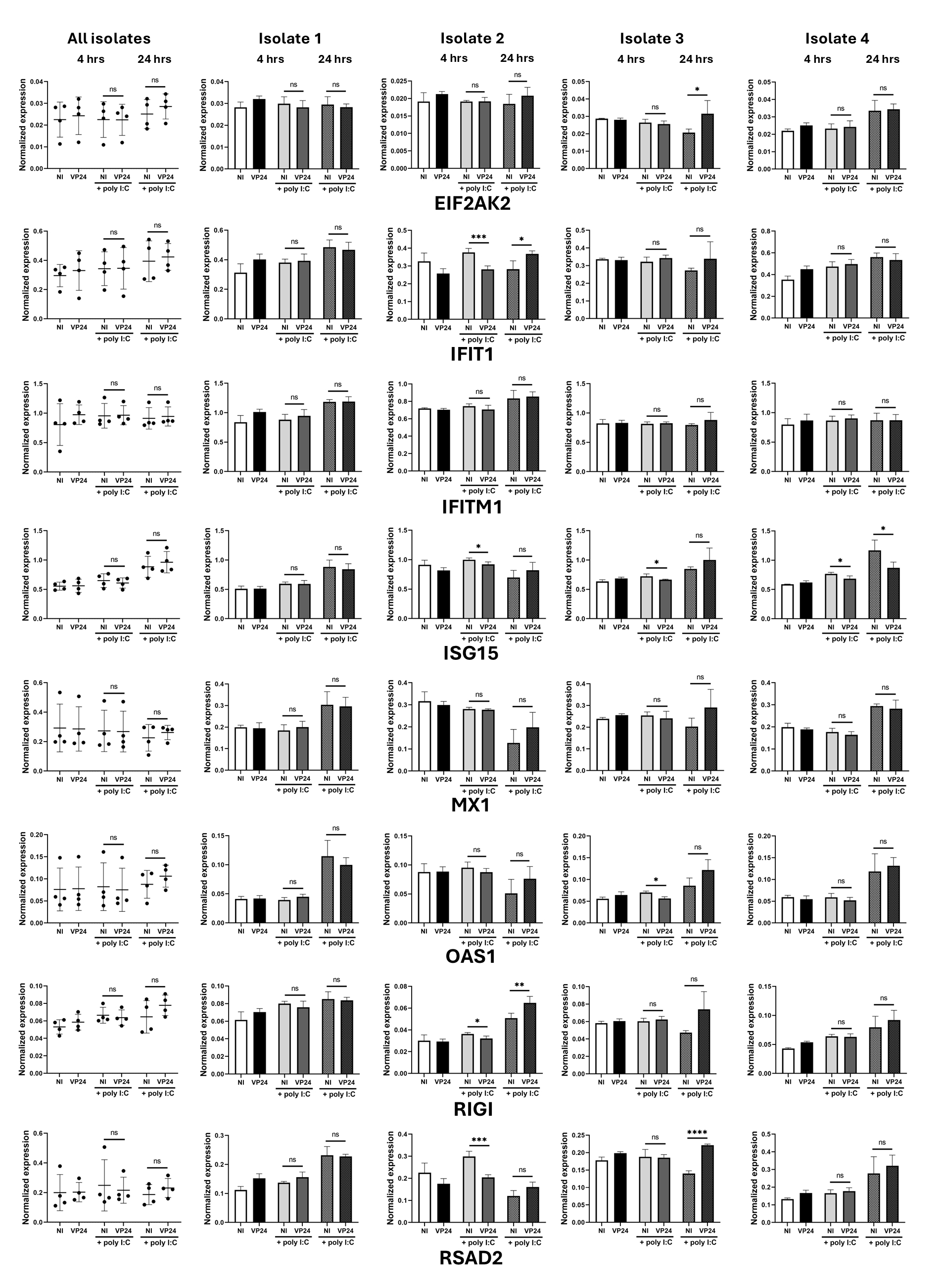
 Figure 3 of
Ashander, Mol Vis 2026; 32:34-48.
Figure 3 of
Ashander, Mol Vis 2026; 32:34-48.