Figure 6. Tasquinimod suppresses angiogenesis-related protein expression in hypoxia-stimulated human retinal capillary endothelial cells
(HRECs). A: Representative western blot images show the expression levels of HIF-1α, VEGF, ICAM-1, and TSP-1 in HRECs under normoxic
or hypoxic conditions, with or without tasquinimod treatment. Protein levels of HIF-1α (B), VEGF (C), ICAM-1 (D), and TSP-1 (E) were quantified and normalized to β-actin. Data are presented as the mean ± SD. ***p < 0.001.
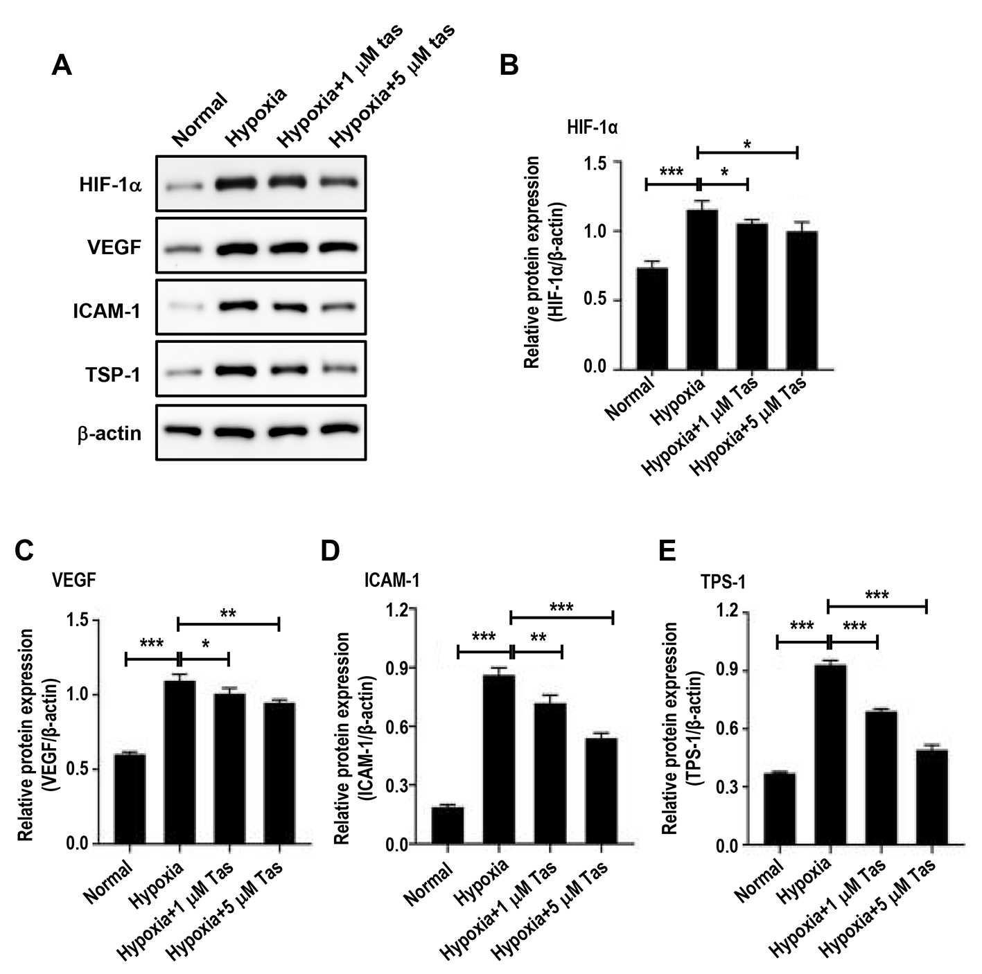
 Figure 6 of
Ji, Mol Vis 2026; 32:325-341.