Figure 4. Hypoxia induces HIF-1α and S100A9 expression in human retinal capillary endothelial cells (HRECs). A: A CCK-8 assay was performed to assess HREC viability after treatment with 100 μM, 200 μM, or 300 μM cobalt chloride (CoCl2)
for 12 h and 24 h. Data are presented as a percentage of the control at 0 h. Quantitative reverse transcription quantitative
polymerase chain reaction analysis was performed to determine HIF-1α (B) and S100A9 (C) mRNA expression under normoxia or 200 μM CoCl2-induced hypoxia, with normalization to β-actin. D: Representative western blots show HIF-1α and S100A9 protein expression in HRECs under normoxic and hypoxic conditions. Protein
levels of HIF-1α (E) and S100A9 (F) are expressed as the ratio of target protein expression to β-actin. Data are presented as the mean ± SD. ***p < 0.001 and
****p < 0.0001.
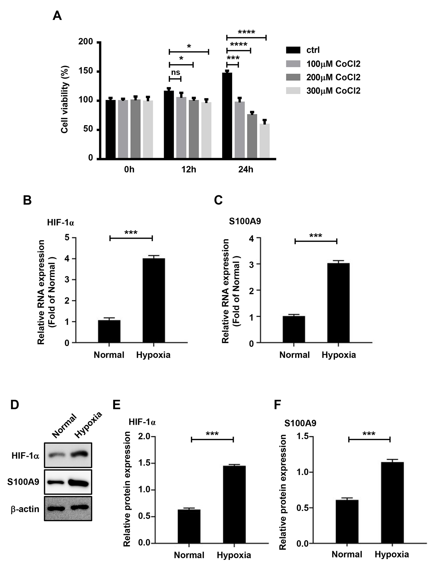
 Figure 4 of
Ji, Mol Vis 2026; 32:325-341.