Figure 4. Bicdl1 KI mice have significantly reduced average thickness of rod photoreceptor outer segment layer in retina cross sections, indicating
shorter outer segments. A-D: Representative retinal sections from 9- and 19-month-old mice immunostained with anti-rhodopsin (white), labeling rod photoreceptor
outer segments. Images were acquired using a 25x objective (NA 1.0). Quantification of the average thickness (area of staining
divided by length of the imaged field) of the rhodopsin-stained outer segments in 9-month-old (E, WT n = 4, KI n = 5) and
19-month-old (F, WT n = 5, KI n = 5) mice is shown for Bicdl1 WT and KI mice in the midperipheral zone. Rho Ab: Rhodopsin antibody, Black squares: WT mice, blue squares: Bicdl1 KI mice. Data are shown as means ± SEM. *: p < 0.05, Scale bar: 100 µm.
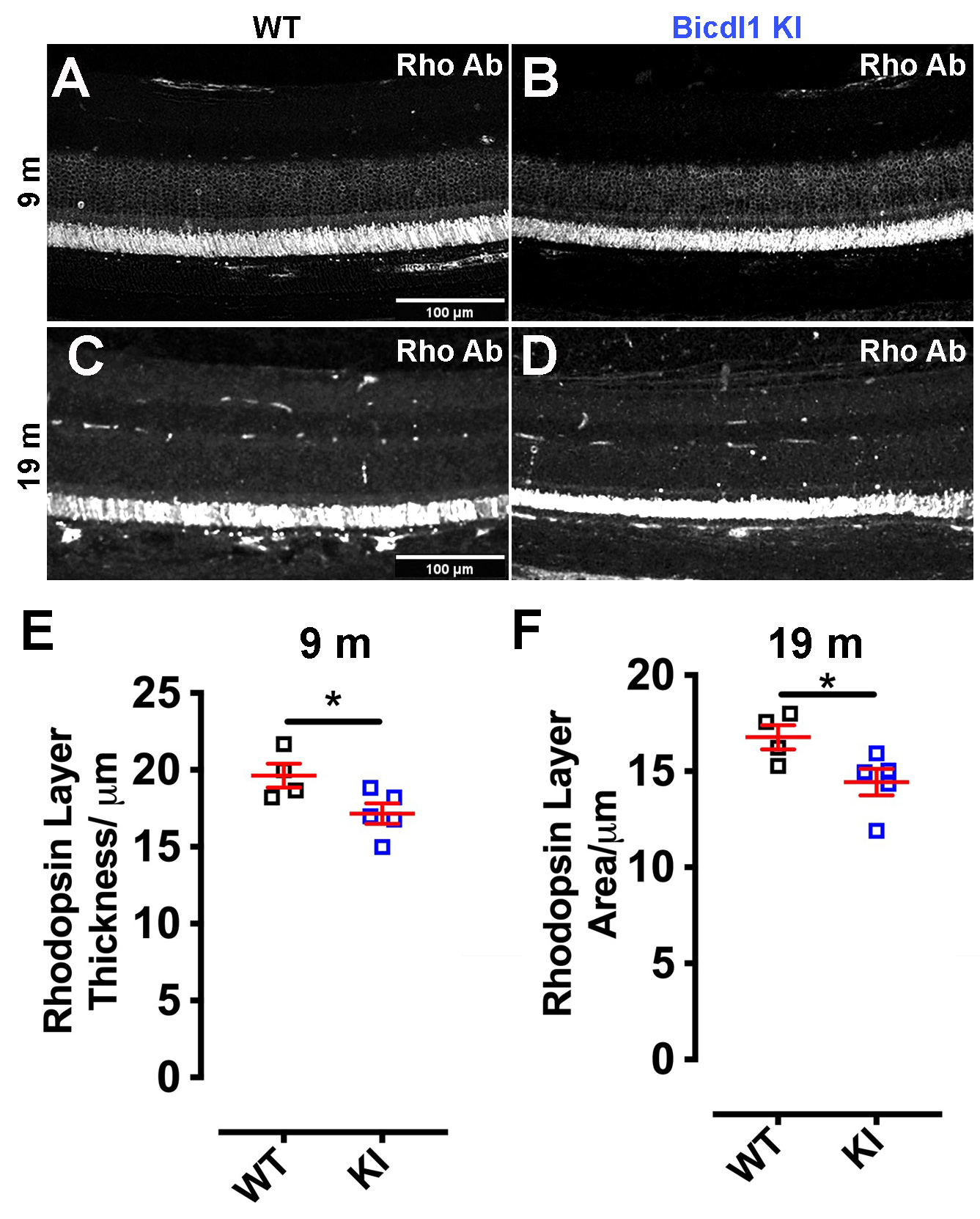
 Figure 4 of
Ulker-Yilmazer, Mol Vis 2026; 32:281-306.
Figure 4 of
Ulker-Yilmazer, Mol Vis 2026; 32:281-306.