Figure 3. Bicdl1 KI mice have a decreased number of photoreceptors and thinning of ONL in H&E-stained retina cross sections. A-D: Representative images of retina sections from 9- and 19-month-old mice stained with H&E. E-H: Quantification of ONL thicknesses (E, G) and ONL nuclei per column (photoreceptor cell body numbers; F, H) at 9 months of age (E, F; WT n = 4, KI n = 5) and 19 months of age (G, H; WT n = 5, KI n = 5). Measurements were taken at 500 µm intervals starting from 300 µm from the optic nerve head (ONH) on
both sides. Black squares: WT mice, blue squares: Bicdl1 KI mice. Data are shown as means ± SEM. # (non-significant trend): p < 0.1, *: p < 0.05, **: p < 0.01, Scale bar: 100 µm.
ONL: outer nuclear layer.
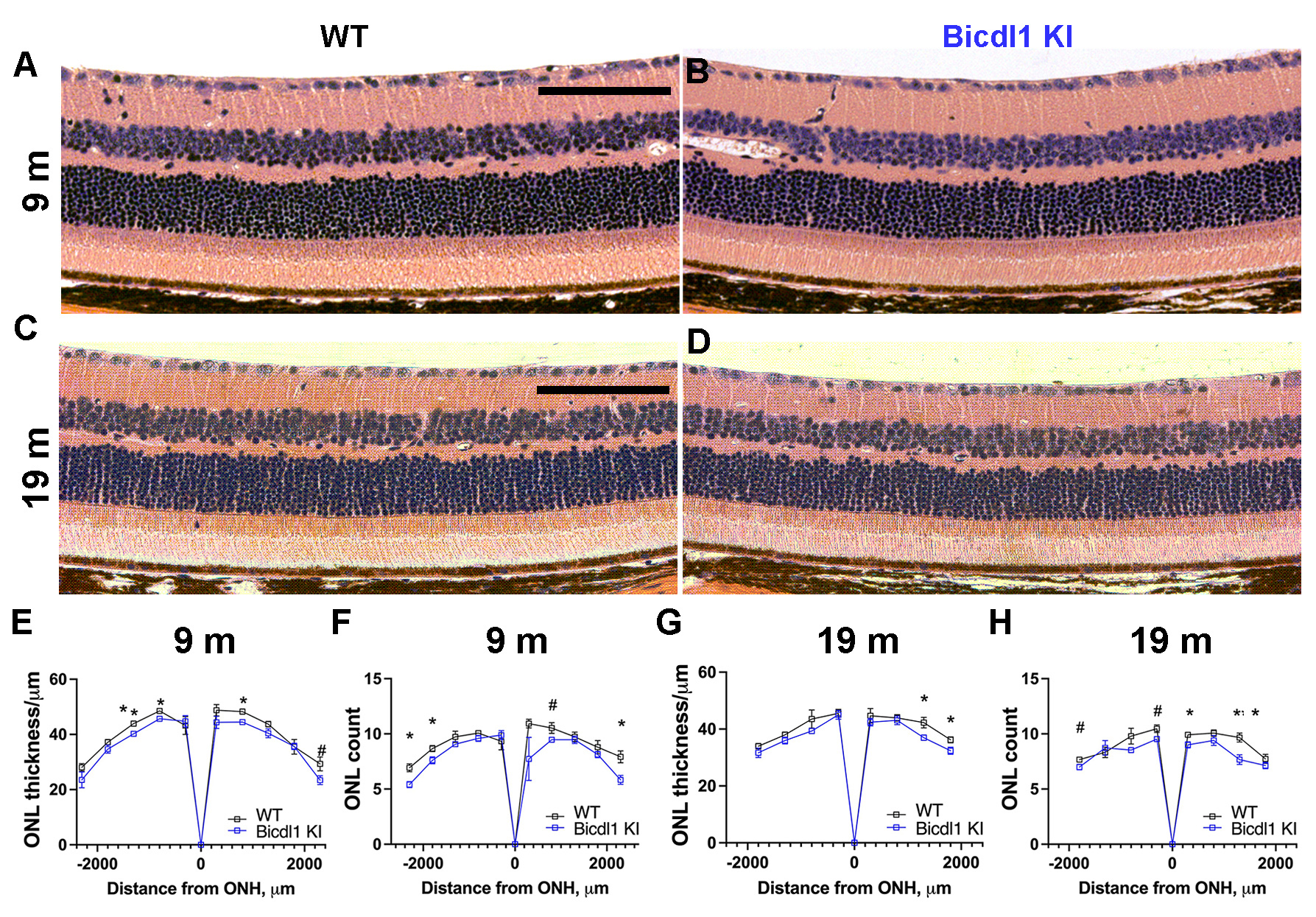
 Figure 3 of
Ulker-Yilmazer, Mol Vis 2026; 32:281-306.
Figure 3 of
Ulker-Yilmazer, Mol Vis 2026; 32:281-306.