Appendix 8 of
Ulker-Yilmazer, Mol Vis 2026; 32:281-306.
Appendix 8 of
Ulker-Yilmazer, Mol Vis 2026; 32:281-306. Appendix 8 of
Ulker-Yilmazer, Mol Vis 2026; 32:281-306.
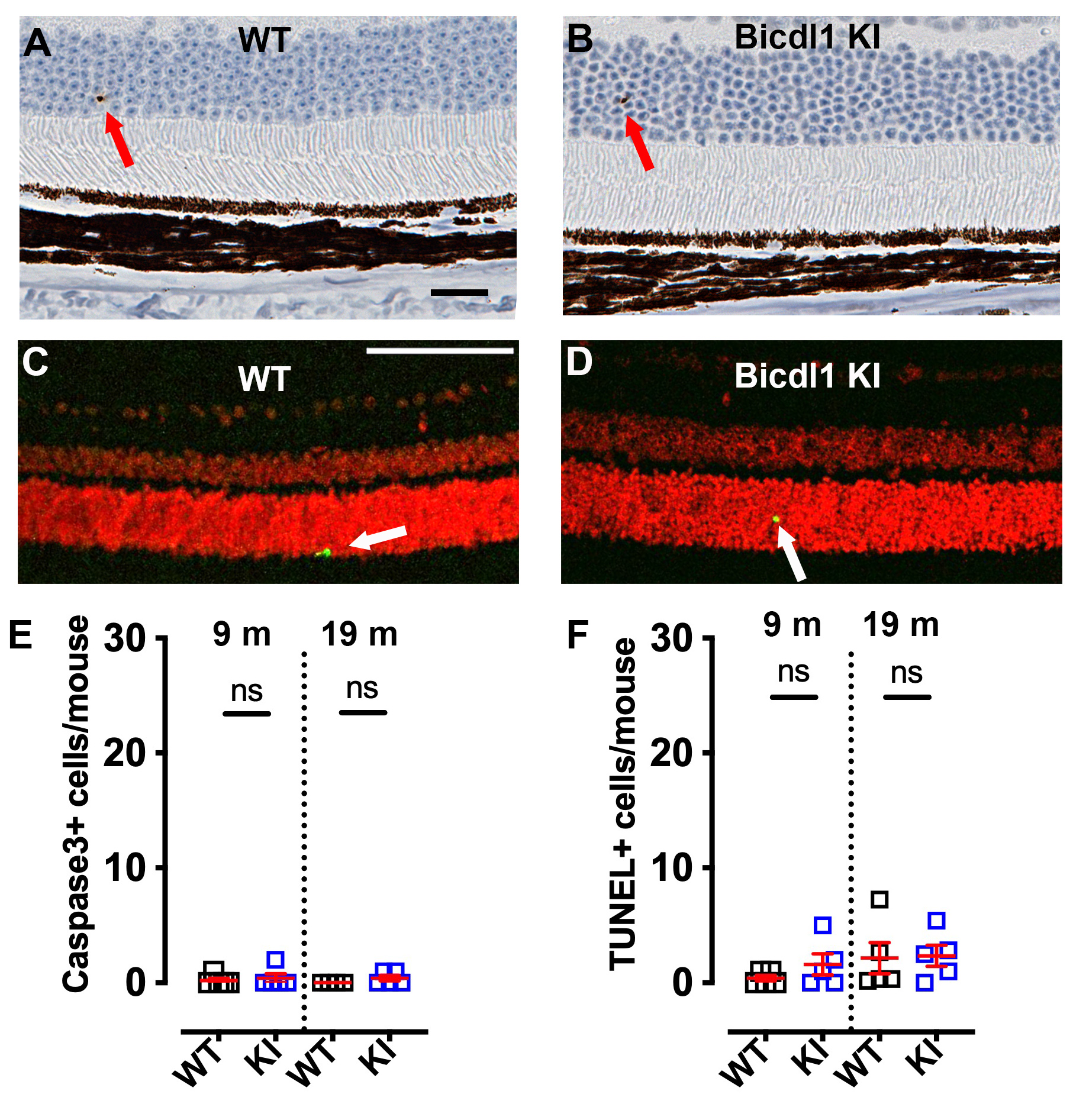
Appendix 8. Supplementary Figure S5: Caspase-3 staining and TUNEL staining of retina sections do not show a significant increase in apoptosis in Bicdl1 KI mice.
To access the data, click or select the words “Appendix 8.” A, B) Representative images of caspase-3 staining in retina sections of 19-month-old WT (A) and Bicdl1 KI (B) . Caspase-3 positive cells are indicated by a red arrow. Scale bar = 20 μm. C, D) Representative images of TUNEL staining in 9-month-old WT (C) and Bicdl1 KI (D) retina sections. TUNEL-positive cells are indicated by a white arrow. Scale bar = 100 μm. E,F) Quantification of caspase-3+ (E) and TUNEL+ (F) cells in retinas of 9- and 19-month-old mice demonstrates that there is a very low level of apoptosis, which was not found to be increased in Bicdl1 KI mice. For caspase staining, n = 5 WT and 5 KI at 9 m, and n = 4 WT and 5 KI at 19 m. For TUNEL staining: n=5 WT and 5 KI at both 9 and 19 months of age. Black squares: WT mice, blue squares: Bicdl1 KI mice. Data are shown as means ± SEM. ns: non-significant.