Appendix 15 of
Ulker-Yilmazer, Mol Vis 2026; 32:281-306.
Appendix 15 of
Ulker-Yilmazer, Mol Vis 2026; 32:281-306. Appendix 15 of
Ulker-Yilmazer, Mol Vis 2026; 32:281-306.
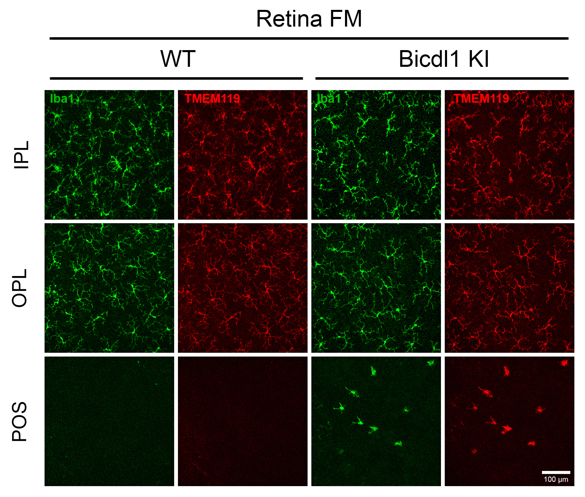
Appendix 15. Supplementary Figure S12: Bicdl1 KI mice display activated microglia in the outer retina.
To access the data, click or select the words “Appendix 15.” Dual immunofluorescence staining of retina flat mounts from 15-month-old WT and Bicdl1 KI mice using antibodies against Iba1 and TMEM119. Confocal images were acquired at the level of the inner plexiform layer (IPL), outer plexiform layer (OPL), and photoreceptor outer segments (POS). Activated microglia accumulation in the POS was observed exclusively in Bicdl1 KI mice. Scale bar = 100 µm.