Appendix 12 of
Ulker-Yilmazer, Mol Vis 2026; 32:281-306.
Appendix 12 of
Ulker-Yilmazer, Mol Vis 2026; 32:281-306. Appendix 12 of
Ulker-Yilmazer, Mol Vis 2026; 32:281-306.
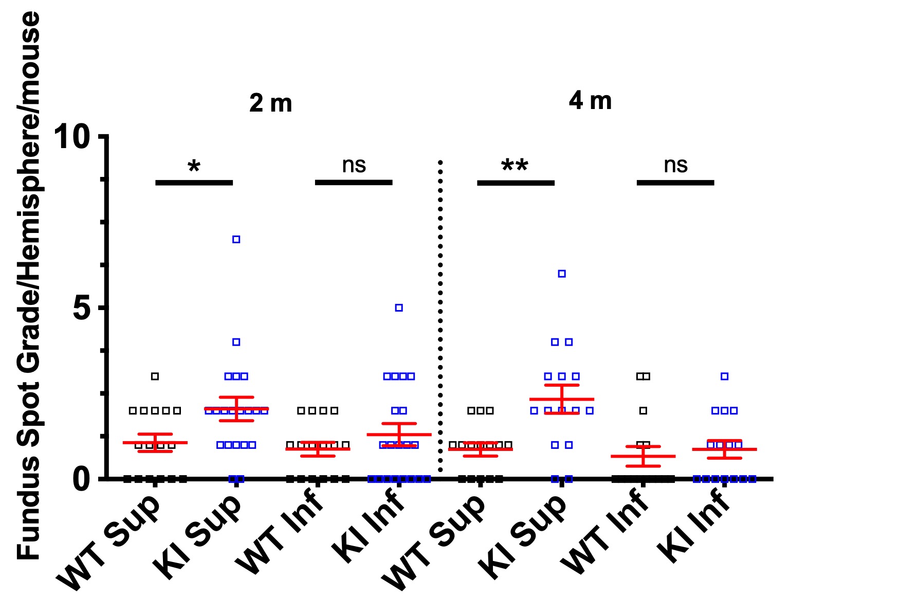
Appendix 12. Supplementary Figure S9: Fundus spots start accumulating in the superior hemiretina of Bicdl1 KI first.
To access the data, click or select the words “Appendix 12.” We could detect a statistically significant increase in fundus spots in the superior hemiretina of Bicdl1 KI compared to WT mice as early as 2 months of age. n = 16 WT and 20 KI at 2 m, and n = 15 WT and 17 KI at 4 m. At these early time points, there was still no significant increase in fundus spots in the inferior retina.